- Mammary Glands
- Fallopian Tubes
- Supporting Ligaments
- Reproductive System
- Gametogenesis
- Placental Development
- Maternal Adaptations
- Menstrual Cycle
- Antenatal Care
- Small for Gestational Age
- Large for Gestational Age
- RBC Isoimmunisation
- Prematurity
- Prolonged Pregnancy
- Multiple Pregnancy
- Miscarriage
- Recurrent Miscarriage
- Ectopic Pregnancy
- Hyperemesis Gravidarum
- Gestational Trophoblastic Disease
- Breech Presentation
- Abnormal lie, Malpresentation and Malposition
- Oligohydramnios
- Polyhydramnios
- Placenta Praevia
- Placental Abruption
- Pre-Eclampsia
- Gestational Diabetes
- Headaches in Pregnancy
- Haematological
- Obstetric Cholestasis
- Thyroid Disease in Pregnancy
- Epilepsy in Pregnancy
- Induction of Labour
- Operative Vaginal Delivery
- Prelabour Rupture of Membranes
- Caesarean Section
- Shoulder Dystocia
- Cord Prolapse
- Uterine Rupture
- Amniotic Fluid Embolism
- Primary PPH
- Secondary PPH
- Psychiatric Disease
- Postpartum Contraception
- Breastfeeding Problems
- Primary Dysmenorrhoea
- Amenorrhoea and Oligomenorrhoea
- Heavy Menstrual Bleeding
- Endometriosis
- Endometrial Cancer
- Adenomyosis
- Cervical Polyps
- Cervical Ectropion
- Cervical Intraepithelial Neoplasia + Cervical Screening
- Cervical Cancer
- Polycystic Ovary Syndrome (PCOS)
- Ovarian Cysts & Tumours
- Urinary Incontinence
- Genitourinary Prolapses
- Bartholin's Cyst
- Lichen Sclerosus
- Vulval Carcinoma
- Introduction to Infertility
- Female Factor Infertility
- Male Factor Infertility
- Female Genital Mutilation
- Barrier Contraception
- Combined Hormonal
- Progesterone Only Hormonal
- Intrauterine System & Device
- Emergency Contraception
- Pelvic Inflammatory Disease
- Genital Warts
- Genital Herpes
- Trichomonas Vaginalis
- Bacterial Vaginosis
- Vulvovaginal Candidiasis
- Obstetric History
- Gynaecological History
- Sexual History
- Obstetric Examination
- Speculum Examination
- Bimanual Examination
- Amniocentesis
- Chorionic Villus Sampling
- Hysterectomy
- Endometrial Ablation
- Tension-Free Vaginal Tape
- Contraceptive Implant
- Fitting an IUS or IUD

Abnormal Fetal lie, Malpresentation and Malposition
Original Author(s): Anna Mcclune Last updated: 1st December 2018 Revisions: 12
- 1 Definitions
- 2 Risk Factors
- 3.2 Presentation
- 3.3 Position
- 4 Investigations
- 5.1 Abnormal Fetal Lie
- 5.2 Malpresentation
- 5.3 Malposition
The lie, presentation and position of a fetus are important during labour and delivery.
In this article, we will look at the risk factors, examination and management of abnormal fetal lie, malpresentation and malposition.
Definitions
- Longitudinal, transverse or oblique
- Cephalic vertex presentation is the most common and is considered the safest
- Other presentations include breech, shoulder, face and brow
- Usually the fetal head engages in the occipito-anterior position (the fetal occiput facing anteriorly) – this is ideal for birth
- Other positions include occipito-posterior and occipito-transverse.
Note: Breech presentation is the most common malpresentation, and is covered in detail here .

Fig 1 – The two most common fetal presentations: cephalic and breech.
Risk Factors
The risk factors for abnormal fetal lie, malpresentation and malposition include:
- Multiple pregnancy
- Uterine abnormalities (e.g fibroids, partial septate uterus)
- Fetal abnormalities
- Placenta praevia
- Primiparity
Identifying Fetal Lie, Presentation and Position
The fetal lie and presentation can usually be identified via abdominal examination. The fetal position is ascertained by vaginal examination.
For more information on the obstetric examination, see here .
- Face the patient’s head
- Place your hands on either side of the uterus and gently apply pressure; one side will feel fuller and firmer – this is the back, and fetal limbs may feel ‘knobbly’ on the opposite side
Presentation
- Palpate the lower uterus (above the symphysis pubis) with the fingers of both hands; the head feels hard and round (cephalic) and the bottom feels soft and triangular (breech)
- You may be able to gently push the fetal head from side to side
The fetal lie and presentation may not be possible to identify if the mother has a high BMI, if she has not emptied her bladder, if the fetus is small or if there is polyhydramnios .
During labour, vaginal examination is used to assess the position of the fetal head (in a cephalic vertex presentation). The landmarks of the fetal head, including the anterior and posterior fontanelles, indicate the position.

Fig 2 – Assessing fetal lie and presentation.
Investigations
Any suspected abnormal fetal lie or malpresentation should be confirmed by an ultrasound scan . This could also demonstrate predisposing uterine or fetal abnormalities.
Abnormal Fetal Lie
If the fetal lie is abnormal, an external cephalic version (ECV) can be attempted – ideally between 36 and 38 weeks gestation.
ECV is the manipulation of the fetus to a cephalic presentation through the maternal abdomen.
It has an approximate success rate of 50% in primiparous women and 60% in multiparous women. Only 8% of breech presentations will spontaneously revert to cephalic in primiparous women over 36 weeks gestation.
Complications of ECV are rare but include fetal distress , premature rupture of membranes, antepartum haemorrhage (APH) and placental abruption. The risk of an emergency caesarean section (C-section) within 24 hours is around 1 in 200.
ECV is contraindicated in women with a recent APH, ruptured membranes, uterine abnormalities or a previous C-section .

Fig 3 – External cephalic version.
Malpresentation
The management of malpresentation is dependent on the presentation.
- Breech – attempt ECV before labour, vaginal breech delivery or C-section
- Brow – a C-section is necessary
- If the chin is anterior (mento-anterior) a normal labour is possible; however, it is likely to be prolonged and there is an increased risk of a C-section being required
- If the chin is posterior (mento-posterior) then a C-section is necessary
- Shoulder – a C-section is necessary
Malposition
90% of malpositions spontaneously rotate to occipito-anterior as labour progresses. If the fetal head does not rotate, rotation and operative vaginal delivery can be attempted. Alternatively a C-section can be performed.
- Usually the fetal head engages in the occipito-anterior position (the fetal occiput facing anteriorly) - this is ideal for birth
If the fetal lie is abnormal, an external cephalic version (ECV) can be attempted - ideally between 36 and 38 weeks gestation.
- Breech - attempt ECV before labour, vaginal breech delivery or C-section
Found an error? Is our article missing some key information? Make the changes yourself here!
Once you've finished editing, click 'Submit for Review', and your changes will be reviewed by our team before publishing on the site.
We use cookies to improve your experience on our site and to show you relevant advertising. To find out more, read our privacy policy .
Privacy Overview
| Cookie | Duration | Description |
|---|---|---|
| cookielawinfo-checkbox-analytics | 11 months | This cookie is set by GDPR Cookie Consent plugin. The cookie is used to store the user consent for the cookies in the category "Analytics". |
| cookielawinfo-checkbox-functional | 11 months | The cookie is set by GDPR cookie consent to record the user consent for the cookies in the category "Functional". |
| cookielawinfo-checkbox-necessary | 11 months | This cookie is set by GDPR Cookie Consent plugin. The cookies is used to store the user consent for the cookies in the category "Necessary". |
| cookielawinfo-checkbox-others | 11 months | This cookie is set by GDPR Cookie Consent plugin. The cookie is used to store the user consent for the cookies in the category "Other. |
| cookielawinfo-checkbox-performance | 11 months | This cookie is set by GDPR Cookie Consent plugin. The cookie is used to store the user consent for the cookies in the category "Performance". |
| viewed_cookie_policy | 11 months | The cookie is set by the GDPR Cookie Consent plugin and is used to store whether or not user has consented to the use of cookies. It does not store any personal data. |
Fetal Presentation, Position, and Lie (Including Breech Presentation)
- Variations in Fetal Position and Presentation |
During pregnancy, the fetus can be positioned in many different ways inside the mother's uterus. The fetus may be head up or down or facing the mother's back or front. At first, the fetus can move around easily or shift position as the mother moves. Toward the end of the pregnancy the fetus is larger, has less room to move, and stays in one position. How the fetus is positioned has an important effect on delivery and, for certain positions, a cesarean delivery is necessary. There are medical terms that describe precisely how the fetus is positioned, and identifying the fetal position helps doctors to anticipate potential difficulties during labor and delivery.
Presentation refers to the part of the fetus’s body that leads the way out through the birth canal (called the presenting part). Usually, the head leads the way, but sometimes the buttocks (breech presentation), shoulder, or face leads the way.
Position refers to whether the fetus is facing backward (occiput anterior) or forward (occiput posterior). The occiput is a bone at the back of the baby's head. Therefore, facing backward is called occiput anterior (facing the mother’s back and facing down when the mother lies on her back). Facing forward is called occiput posterior (facing toward the mother's pubic bone and facing up when the mother lies on her back).
Lie refers to the angle of the fetus in relation to the mother and the uterus. Up-and-down (with the baby's spine parallel to mother's spine, called longitudinal) is normal, but sometimes the lie is sideways (transverse) or at an angle (oblique).
For these aspects of fetal positioning, the combination that is the most common, safest, and easiest for the mother to deliver is the following:
Head first (called vertex or cephalic presentation)
Facing backward (occiput anterior position)
Spine parallel to mother's spine (longitudinal lie)
Neck bent forward with chin tucked
Arms folded across the chest
If the fetus is in a different position, lie, or presentation, labor may be more difficult, and a normal vaginal delivery may not be possible.
Variations in fetal presentation, position, or lie may occur when
The fetus is too large for the mother's pelvis (fetopelvic disproportion).
The uterus is abnormally shaped or contains growths such as fibroids .
The fetus has a birth defect .
There is more than one fetus (multiple gestation).

Position and Presentation of the Fetus
Toward the end of pregnancy, the fetus moves into position for delivery. Normally, the presentation is vertex (head first), and the position is occiput anterior (facing toward the pregnant person's spine) and with the face and body angled to one side and the neck flexed. Variations in fetal presentations include face, brow, breech, and shoulder. Occiput posterior position (facing forward, toward the mother's pubic bone) is less common than occiput anterior position (facing backward, toward the mother's spine). |
Variations in Fetal Position and Presentation
Some variations in position and presentation that make delivery difficult occur frequently.
Occiput posterior position
In occiput posterior position (sometimes called sunny-side up), the fetus is head first (vertex presentation) but is facing forward (toward the mother's pubic bone—that is, facing up when the mother lies on her back). This is a very common position that is not abnormal, but it makes delivery more difficult than when the fetus is in the occiput anterior position (facing toward the mother's spine—that is facing down when the mother lies on her back).
When a fetus faces up, the neck is often straightened rather than bent,which requires more room for the head to pass through the birth canal. Delivery assisted by a vacuum device or forceps or cesarean delivery may be necessary.
Breech presentation
In breech presentation, the baby's buttocks or sometimes the feet are positioned to deliver first (before the head).
When delivered vaginally, babies that present buttocks first are more at risk of injury or even death than those that present head first.
The reason for the risks to babies in breech presentation is that the baby's hips and buttocks are not as wide as the head. Therefore, when the hips and buttocks pass through the cervix first, the passageway may not be wide enough for the head to pass through. In addition, when the head follows the buttocks, the neck may be bent slightly backwards. The neck being bent backward increases the width required for delivery as compared to when the head is angled forward with the chin tucked, which is the position that is easiest for delivery. Thus, the baby’s body may be delivered and then the head may get caught and not be able to pass through the birth canal. When the baby’s head is caught, this puts pressure on the umbilical cord in the birth canal, so that very little oxygen can reach the baby. Brain damage due to lack of oxygen is more common among breech babies than among those presenting head first.
In a first delivery, these problems may occur more frequently because a woman’s tissues have not been stretched by previous deliveries. Because of risk of injury or even death to the baby, cesarean delivery is preferred when the fetus is in breech presentation, unless the doctor is very experienced with and skilled at delivering breech babies or there is not an adequate facility or equipment to safely perform a cesarean delivery.
Breech presentation is more likely to occur in the following circumstances:
Labor starts too soon (preterm labor).
The uterus is abnormally shaped or contains abnormal growths such as fibroids .
Other presentations
In face presentation, the baby's neck arches back so that the face presents first rather than the top of the head.
In brow presentation, the neck is moderately arched so that the brow presents first.
Usually, fetuses do not stay in a face or brow presentation. These presentations often change to a vertex (top of the head) presentation before or during labor. If they do not, a cesarean delivery is usually recommended.
In transverse lie, the fetus lies horizontally across the birth canal and presents shoulder first. A cesarean delivery is done, unless the fetus is the second in a set of twins. In such a case, the fetus may be turned to be delivered through the vagina.

Copyright © 2024 Merck & Co., Inc., Rahway, NJ, USA and its affiliates. All rights reserved.
- Cookie Preferences

Website maintenance is scheduled for Saturday, September 7, and Sunday, September 8. Short disruptions may occur during these days.

MONICA PREBOTH
Am Fam Physician. 2000;62(5):1184-1188
The Committee on Practice Bulletins–Obstetrics of the American College of Obstetricians and Gynecologists (ACOG) has developed clinical management guidelines on antepartum fetal surveillance. According to the committee, the goal of antepartum fetal surveillance is to prevent fetal death. The techniques of antepartum fetal surveillance, which are based on the assessment of fetal heart rate patterns, have been in clinical use for nearly 30 years. These guidelines, which replace Technical Bulletin No. 188 issued in January 1994, appear in the October 1999 issue of Obstetrics and Gynecology .
Techniques of Antepartum Fetal Surveillance
Several techniques for antepartum fetal surveillance currently in use are discussed in the ACOG bulletin. These include fetal movement assessment, nonstress test, contraction stress test, fetal biophysical profile, modified biophysical profile and umbilical artery Doppler velocimetry.
FETAL MOVEMENT ASSESSMENT
Fetal movement assessment occurs when the mother perceives a diminution in fetal movement. The mother counts fetal “kicks” as a means of antepartum fetal surveillance. The optimal number of movements and the ideal duration for counting movements have not been determined; however, numerous protocols have been reported and appear to be acceptable.
CONTRACTION STRESS TEST
The contraction stress test is based on the response of the fetal heart rate to uterine contractions. It is believed that fetal oxygenation will be transiently worsened by uterine contractions. In the fetus with suboptimal oxygenation, the resulting intermittent worsening in oxygenation will, in turn, lead to the fetal heart rate pattern of late decelerations. Uterine contractions also may provoke or accentuate a pattern of variable decelerations caused by fetal umbilical cord compression, which in some cases is associated with oligohydramnios.
The contraction stress test is interpreted by the presence or absence of late fetal heart rate decelerations, which are defined as decelerations that reach their nadir after the peak of the contraction and that usually persist beyond the end of the contraction. The results of the contraction stress test are categorized in the ACOG bulletin as follows:
Negative . No late or significant variable decelerations.
Positive . Late decelerations following 50 percent or more of contractions (even if the contraction frequency is fewer than three in 10 minutes).
Equivocal - suspicious . Intermittent late decelerations or significant variable decelerations.
Equivocal - hyperstimulatory . Fetal heart rate decelerations that occur in the presence of contractions that are more frequent than every two minutes or last longer than 90 seconds.
Unsatisfactory . Fewer than three contractions in 10 minutes or a tracing that is not interpretable.
Relative contraindications to the contraction stress test usually include conditions that are associated with an increased risk of preterm labor and delivery, uterine rupture or uterine bleeding. According to ACOG, these conditions include the following:
Preterm labor or certain patients at high risk of preterm labor.
Preterm membrane rupture.
History of extensive uterine surgery or classic cesarean delivery.
Known placenta previa.
NONSTRESS TEST
In the nonstress test, the heart rate of the fetus that is not acidotic or neurologically depressed will temporarily accelerate with fetal movement. Heart rate reactivity is believed to be a good indicator of normal fetal autonomic function. Loss of reactivity is commonly associated with a fetal sleep cycle but may result from any cause of central nervous system depression, including fetal acidosis.
Results of nonstress tests are classified as reactive or nonreactive. Various definitions of reactivity have been used. Most commonly, the nonstress test is considered reactive, or normal, if there are two or more fetal heart rate accelerations within a 20-minute period, with or without fetal movement discernible by the woman, according to ACOG. The nonreactive stress test lacks sufficient fetal heart rate accelerations over a 40-minute period. The nonstress test of the neurologically healthy preterm fetus is frequently nonreactive—from 24 to 28 weeks of gestation, up to 50 percent of nonstress tests may not be reactive, and from 28 to 32 weeks of gestation, 15 percent of nonstress tests are not reactive.
BIOPHYSICAL PROFILE
The biophysical profile discussed in the ACOG bulletin is a nonstress test plus four observations made by real-time ultrasonography. The five components of the biophysical profile are as follows: (1) nonstress test; (2) fetal breathing movements (one or more episodes of rhythmic fetal breathing movements of 30 seconds or more within 30 minutes); (3) fetal movement (three or more discrete body or limb movements within 30 minutes); (4) fetal tone (one or more episodes of extension of a fetal extremity with return to flexion, or opening or closing of a hand; and (5) determination of the amniotic fluid volume (a single vertical pocket of amniotic fluid exceeding 2 cm is considered evidence of adequate amniotic fluid).
Each of the components is given a score of 2 (normal or present as defined previously) or 0 (abnormal, absent or insufficient). A composite score of 8 or 10 is normal, a score of 6 is equivocal and a score of 4 or less is abnormal. In the presence of oligohydramnios, further evaluation is warranted regardless of the composite score.
MODIFIED BIOPHYSICAL PROFILE
During the late second or third trimester, amniotic fluid reflects fetal urine production. Placental dysfunction may cause diminished fetal renal perfusion, which can lead to oligohydramnios. Therefore, assessment of amniotic fluid volume can be used to evaluate long-term uteroplacental function. This led to the development of the modified biophysical profile.
The modified biophysical profile combines the non-stress test with the amniotic fluid index, which is the sum of measurements of the deepest cord-free amniotic fluid pocket in each of the abdominal quadrants, as an indicator of long-term function of the placenta. An amniotic fluid index of more than 5 cm is thought to be an adequate volume of amniotic fluid. The modified biophysical profile is considered normal if the nonstress test is reactive and the amniotic fluid index is greater than 5 cm and abnormal if the nonstress test is nonreactive or the amniotic fluid index is 5 cm or less.
UMBILICAL ARTERY DOPPLER VELOCIMETRY
Doppler ultrasonography is used to assess the hemodynamic components of vascular impedence. Umbilical artery Doppler flow velocimetry has been adapted as a fetal surveillance technique because it is believed that flow velocity waveforms in the umbilical artery of fetuses with normal growth differ from those of fetuses with growth restriction. The umbilical flow velocity waveform of a normally growing fetus has high-velocity diastolic flow, while in cases of intrauterine growth restriction, the umbilical artery diastolic flow is diminished. With extreme intrauterine growth restriction, the flow may be absent or even reversed. There is a high perinatal mortality rate among such pregnancies.
Indications for Antepartum Fetal Surveillance
The results of antepartum fetal surveillance have not definitively demonstrated improved perinatal outcome. Therefore, all indications for antepartum testing should be considered somewhat relative. Usually, antepartum fetal surveillance is used in pregnancies with a high risk of antepartum fetal death. Some of the conditions in which testing is appropriate include the following:
Maternal conditions: antiphospholipid syndrome, poorly controlled hyperthyroidism, hemoglobinopathies such as hemoglobin SS, SC or S-thalassemia, cyanotic heart disease, systemic lupus erythematosus, chronic renal disease, type 1 diabetes mellitus and hypertensive disorders.
Pregnancy-related conditions : pregnancy-induced hypertension, decreased fetal movement, oligohydramnios, polyhydramnios, intrauterine growth restriction, post-term pregnancy, moderate to severe isoimmunization, previous fetal demise (unexplained or recurrent risk) and multiple gestation with significant growth discrepancy.
Recommendations
The following ACOG recommendations are based on limited or inconsistent scientific evidence (Level B):
Women at high risk for stillbirth should undergo antepartum fetal surveillance using the nonstress test, contraction stress test, biophysical profile or modified biophysical profile.
Initiation of testing at 32 to 34 weeks of gestation is appropriate for most pregnancies that are at increased risk of stillbirth. In pregnancies with multiple or particularly worrisome high-risk conditions, testing may be initiated as early as 26 to 28 weeks of gestation.
When the clinical condition that prompted testing persists, a reassuring test should be repeated weekly or, depending on the test used and the presence of certain high-risk conditions, twice weekly until delivery. Any significant deterioration in fetal activity requires fetal reevaluation, regardless of the amount of time that has elapsed since the last test.
An abnormal nonstress test or modified biophysical profile usually should be further evaluated by a contraction stress test or a full biophysical profile. Subsequent management should then be predicated on the results of the contraction stress test or biophysical profile, the gestational age, the degree of oligohydramnios (if assessed) and the maternal condition.
Oligohydramnios, defined as no ultrasonographically measurable vertical pocket of amniotic fluid greater than 2 cm or an amniotic fluid index of 5 cm or less, requires (depending on the degree of oligohydramnios, the gestational age and the maternal clinical condition) delivery, or close maternal or fetal surveillance.
In the absence of obstetric contraindications, delivery of the fetus with an abnormal test result often may be attempted by induction of labor with continuous monitoring of the fetal heart rate and contractions. If repetitive late decelerations are observed, cesarean delivery generally is indicated.
Recent, normal antepartum fetal test results should not preclude the use of intrapartum fetal monitoring.
Umbilical artery Doppler velocimetry seems to benefit only pregnancies complicated by intrauterine growth restriction. If used in this setting, decisions regarding timing of delivery should be made using a combination of information from the Doppler ultrasonography and other tests of fetal well-being, along with careful monitoring of maternal status.
Middle cerebral artery Doppler velocimetry should be considered an investigational approach to antepartum fetal surveillance.
Coverage of guidelines from other organizations does not imply endorsement by AFP or the AAFP.
This series is coordinated by Michael J. Arnold, MD, Assistant Medical Editor.
A collection of Practice Guidelines published in AFP is available at https://www.aafp.org/afp/practguide .
Continue Reading
More in afp, more in pubmed.
Copyright © 2000 by the American Academy of Family Physicians.
This content is owned by the AAFP. A person viewing it online may make one printout of the material and may use that printout only for his or her personal, non-commercial reference. This material may not otherwise be downloaded, copied, printed, stored, transmitted or reproduced in any medium, whether now known or later invented, except as authorized in writing by the AAFP. See permissions for copyright questions and/or permission requests.
Copyright © 2024 American Academy of Family Physicians. All Rights Reserved.
Fetal Presentation, Position, and Lie (Including Breech Presentation)
- Key Points |
Abnormal fetal lie or presentation may occur due to fetal size, fetal anomalies, uterine structural abnormalities, multiple gestation, or other factors. Diagnosis is by examination or ultrasonography. Management is with physical maneuvers to reposition the fetus, operative vaginal delivery , or cesarean delivery .
Terms that describe the fetus in relation to the uterus, cervix, and maternal pelvis are
Fetal presentation: Fetal part that overlies the maternal pelvic inlet; vertex (cephalic), face, brow, breech, shoulder, funic (umbilical cord), or compound (more than one part, eg, shoulder and hand)
Fetal position: Relation of the presenting part to an anatomic axis; for vertex presentation, occiput anterior, occiput posterior, occiput transverse
Fetal lie: Relation of the fetus to the long axis of the uterus; longitudinal, oblique, or transverse
Normal fetal lie is longitudinal, normal presentation is vertex, and occiput anterior is the most common position.
Abnormal fetal lie, presentation, or position may occur with
Fetopelvic disproportion (fetus too large for the pelvic inlet)
Fetal congenital anomalies
Uterine structural abnormalities (eg, fibroids, synechiae)
Multiple gestation
Several common types of abnormal lie or presentation are discussed here.

Transverse lie
Fetal position is transverse, with the fetal long axis oblique or perpendicular rather than parallel to the maternal long axis. Transverse lie is often accompanied by shoulder presentation, which requires cesarean delivery.
Breech presentation
There are several types of breech presentation.
Frank breech: The fetal hips are flexed, and the knees extended (pike position).
Complete breech: The fetus seems to be sitting with hips and knees flexed.
Single or double footling presentation: One or both legs are completely extended and present before the buttocks.
Types of breech presentations
|
Breech presentation makes delivery difficult ,primarily because the presenting part is a poor dilating wedge. Having a poor dilating wedge can lead to incomplete cervical dilation, because the presenting part is narrower than the head that follows. The head, which is the part with the largest diameter, can then be trapped during delivery.
Additionally, the trapped fetal head can compress the umbilical cord if the fetal umbilicus is visible at the introitus, particularly in primiparas whose pelvic tissues have not been dilated by previous deliveries. Umbilical cord compression may cause fetal hypoxemia.

Predisposing factors for breech presentation include
Preterm labor
Uterine abnormalities
Fetal anomalies
If delivery is vaginal, breech presentation may increase risk of
Umbilical cord prolapse
Birth trauma
Perinatal death

Face or brow presentation
In face presentation, the head is hyperextended, and position is designated by the position of the chin (mentum). When the chin is posterior, the head is less likely to rotate and less likely to deliver vaginally, necessitating cesarean delivery.
Brow presentation usually converts spontaneously to vertex or face presentation.
Occiput posterior position
The most common abnormal position is occiput posterior.
The fetal neck is usually somewhat deflexed; thus, a larger diameter of the head must pass through the pelvis.
Progress may arrest in the second phase of labor. Operative vaginal delivery or cesarean delivery is often required.
Position and Presentation of the Fetus
Toward the end of pregnancy, the fetus moves into position for delivery. Normally, the presentation is vertex (head first), and the position is occiput anterior (facing toward the pregnant patient's spine) with the face and body angled to one side and the neck flexed. Abnormal presentations include face, brow, breech, and shoulder. Occiput posterior position (facing toward the pregnant patient's pubic bone) is less common than occiput anterior position. |
If a fetus is in the occiput posterior position, operative vaginal delivery or cesarean delivery is often required.
In breech presentation, the presenting part is a poor dilating wedge, which can cause the head to be trapped during delivery, often compressing the umbilical cord.
For breech presentation, usually do cesarean delivery at 39 weeks or during labor, but external cephalic version is sometimes successful before labor, usually at 37 or 38 weeks.
Copyright © 2024 Merck & Co., Inc., Rahway, NJ, USA and its affiliates. All rights reserved.
- Cookie Preferences
Phone 6228 6890
Address 18 Main Road, Moonah, Tasmania
Women's Imaging
20 week scan (morphology scan).
Our highly trained sonographers take a detailed look at the anatomy of your baby including the heart and the brain.
This is the main ultrasound scan that is obtained during the pregnancy.
By 20 weeks most of your baby has developed such that screening of the organs is possible to assess for abnormalities. This scan can be done anytime between 18-22 weeks of your pregnancy but ideally around the 20 week mark.
The scan can take a minimum of 45 minutes and sometimes up to an hour.
At this scan the different parts of the baby will be measured to confirm that there has been satisfactory growth in line with that is expected from the due date.
Multiple images of different organs will be taken to confirm that the development is progressing as expected.
While most of the imaging is targeted to the baby, the placenta, umbilical cord and amniotic fluid are also imaged. The blood flow to the uterus will be assessed with Doppler measurements.
Often the sex of the baby can be identified. Please let us know if you would like to know this.
Occasionally not all of the baby can be assessed at one examination. This may be because of the position of the baby. A repeat examination is sometimes required. This will be part of the original examination fee and a booking will be made for you prior to leaving the practice. Ideally we will try and complete the entire scan on the day but sometimes bub doesn’t want to show us everything! Your baby can be comfy in one spot for the entire scan!
What will happen on the day
When you arrive for your scan you will be asked to fill out a form about your pregnancy to date.
The sonographer will then take you through to the scanning room.
You will be asked to lie on the table and expose your tummy. A towel will be tucked into your pants to limit spread of the gel onto your clothes.
Clear gel is applied to your tummy and the sonographer moves the probe over your tummy recording images.
Please come with a partially filled bladder which will make it easier to obtain images of your baby.
Ultrasound at 18 weeks
We will take measurements of your baby and check your placenta and your cervix.
Occasionally a vaginal scan is also performed to give us a better view of your baby or if you have a low-lying placenta . You will be able to watch the entire scan on our wall-mounted plasma screen monitors!
In this scan, we like your baby to move around a little (not alot!) so we can scan everything we want to see.
All of our sonographers are accredited with the Fetal Medicine Foundation. Each sonographer’s scanning style is different. Some like to talk you through the scan whilst others like to stay silent as that helps them concentrate on your baby. The sonographer will explain this to you before the scan. Getting a detailed look at your baby’s anatomy is our number one priority.
Once the scan is completed, the sonographer will leave the room and the results of the scan will be reviewed by our radiologist or on-site obstetrician. If there is any cause for concern, they will come and speak to you on the day. Occasionally your baby is just too comfortable in one spot and we are unable to complete the full scan. This means that you may need to come back again for us to review the parts we haven’t seen.
You will receive a text message within 24 hours of your appointment with a link to access your images through our online portal. Your images will be available to view within 3 days of your appointment.
Ultrasound has been used for many years in pregnancy and is regarded as safe to you and your baby.
Sometimes we need to do a vaginal scan. If you are allergic to latex prior to the vaginal scan or you don’t know then a latex-free cover will be used on the probe.
Occasionally there is some discomfort from probe pressure on your bladder or from the vaginal probe manipulation. If this is extremely painful please let us know.
Unfortunately a normal ultrasound does not guarantee a normal baby. There are regrettably many problems that cannot be determined through imaging. The baby still has 20 weeks to continue to grow and develop.
You will be able to see your developing baby
We are able to assess the heart and brain in great detail.
If there is anything that we are concerned about from the scan we will inform you on the day. This is very reassuring for you and your baby.
What is an Ultrasound scan?
An ultrasound scan uses high-frequency sound waves to create images of the inside of the body and your baby. Sound waves are used instead of radiation which makes them safe.
Why have a 20 week scan at Women’s Imaging?
Our sonographers are trained to the highest standards to perform these specialist measurements of you and your baby. They hold Certificates in Competence from the Fetal Medicine Foundation which is recognised by the Royal Australian and New Zealand College of Obstetricians and Gynaecologists. We take detailed measurements of your baby. All of our scans are monitored by our in-house radiologist or obstetrician.
How long will it take?
The 20 week scan takes approximately 45 minutes but please give yourself lots of time. Occasionally we can’t see/scan everything because of your baby’s position or the position of your placenta. Your own BMI also affects how well we can see your baby on ultrasound. Sometimes you have to rebook for another day so we can complete the scan fully.
How should I prepare for the scan?
You should arrive for your scan with a partially filled bladder. Remember to bring your referral form with you.
Important things to know
- The scan will take about 45 minutes.
- Often the sex of the baby can be identified, please let us know if you would like to know this.
- Our highly accredited sonographers work hard at checking all of your baby’s anatomy including the heart and brain. Please be aware that some sonographers like to scan for the most part in silence as it helps them concentrate on your baby. They will explain to you their scanning style at the start of the scan.
would you like to make an appointment?
great! Just tell us a little bit about yourself and we'll book you in at a time that suits you!
- share on facebook
- share on twitter
- share on google+
- share by email
This appointment cannot be made online. Please contact our friendly reception team on 03 6228 6890 to assist with booking your appointment.
Unfortunately due to COVID-19 safety precautions we are unable to perform BabyView scans at this time. We apologise for any inconvenience. 3D images are still performed as part of our other obstetric scans.
to view your images online
I'm a patient
I'm a doctor.
Download FREE Practo app
Get ₹200 HealthCash
- Consult with a doctor
- Pregnancy and Infertility
Pregnancy. Position change with time?

Answers ( 7 )
Like the answers? Consult privately with the doctor of your choice
Didn't find the answer you are looking for?
Talk to experienced gynaecologist online and get your health questions answered in just 5 minutes.

Articles you may like
Hypertension in Pregnancy
Dr.Pallavi Vasal
Hypertension is defined as high blood pressure (B.P). A person has high blood pressure when the systolic pressure (the t ... Read more
Pregnancy & Oral Health
Dr.Sameer Bhandari
Pregnancy brings about lot of changes in the Mother's body , gums & teeth are the most common victims of negligence ... Read more
6 Useful Tips to Prevent Complications in Pregnancy
Dr.Mohan Pawar
1. PRE CONCEPTIONAL CONSULTATION - Planned pregnancy always has a better outcome than unplanned pregnancy. So have a pr ... Read more
All About Ectopic Pregnancy
Dr.Vaishali Sharma
What is an Ectopic pregnancy?Normally pregnancy occurs inside the womb. Rarely, pregnancy can be implanted in tissues ou ... Read more
Pregnancy Induced Hypertension
Dr.Bimla Bansal
Pregnancy Induced Hypertension, also called PIH, is very important to know about. If not taken care of in time, it can l ... Read more
Pregnancy: Understanding High Risk Pregnancies
High risk pregnancies, as the name suggests, is the condition in which the pregnancy is complicated in some way as compa ... Read more
Doubts about periods or pregnancy?
Consult with a gynaecologist online
- Private & confidential
- Share reports
- Free follow-ups
The question asked on this page is a free question. You can ask a free health question by downloading the Practo app.
Tell us what's troubling you
Periods being irregular.
Consult Now
Vaginal infection troubling you?
Trying to conceive, too many pregnancy doubts, related questions, anomaly scan after 21 week, is any scan required after anomaly scan, anomaly scan, cephalic presentation, mid preg. anomaly scan, query related to anomaly scan report, is everything normal in my scan, b hcg level drop at 11 weeks.
Fetal Weight Percentile Calculator
What percentile is my baby, and what does it mean, how to use the fetal weight percentile calculator, fetal growth chart, how to calculate the fetal weight percentile.
Our fetal weight percentile calculator computes your child's growth and compares it to the general population . Our BPD, HC, AC, and FL calculator uses all the necessary ultrasound fetal measurements to compute your child's weight at a given week of the pregnancy.
We use the Hadlock equation and WHO multinational growth charts . 👶👶🏾👶🏽👶🏻
Want to know more? Follow the article below to discover fetal weight percentile chart by week , and details of our baby weight predictor calculator. We'll also cover the baby percentiles during pregnancy as well as the SGA and LGA definitions.
We use percentiles to measure values that are extremely variable in the population – especially in children. For example, we can't give a precise value of height that a healthy 2-year-old boy should achieve – it all depends on his individual and genetic characteristics . 🔬
However, doctors love all the hard data and numbers that help them make critical clinical decisions. They needed a tool that allowed quick and precise evaluation of their patients.
So how did we tackle this problem? 🤔
Percentiles are a great solution! Instead of giving us a precise number, they inform us about the range in which your baby's height or weight should fall. Thanks to percentiles, we can tell how your child's health looks when compared to the general population and detect any significant abnormalities.
When we talk about fetal weight abnormalities, two most important definitions come to mind:
- Small for the gestational age (age of the pregnancy, counted from the conception; to calculate it, you can use gestational age calculator ); and
- Large for the gestational age.
💡 SGA – Small for Gestational Age babies are all babies that fall below the 10th percentile of the population, when it comes to their EFW percentile. It means that over 90% of the population grew larger than they did.
💡 LGA – Large for Gestational Age babies exceed the 90th percentile of their Estimated Fetal Weight. It means that only 10% of the population is larger than them.
These two explanations are based on an Estimated Fetal Weight (EFW) calculations that use the ultrasound technique to evaluate a baby's weight inside its mother's uterus.
To estimate the fetal weight, we use the following measurements :
Abdominal circumference – circumference of the baby's belly, measured at the liver's height. (Advanced: an umbilical portion of the left portal vein should be in the center of the abdomen.)
Head circumference - measured at the point where specific brain structures are visible. (Advanced: plane that traverses the thalami and cavum septum pellucidum.) To check where the newborn's head circumference places amongst their peers, use head circumference percentile calculator .
Biparietal diameter – length of a distance between two parietal bones of a baby's skull.
Femur length – a length of the baby's thigh bone.
Our fetal growth calculator uses all four of the enumerated measurements; additionally, we can also measure the humerus length .
💡 We could also measure the estimated fetal weight by assessing the fundal height – this method, however, is not as accurate. To learn more about the concept, visit fundal height calculator .
Our estimated fetal weight percentile calculator needs just a few easy steps:
Enter the gender of your baby
Be specific; if you already know the baby's gender, reveal it. 🎉
Enter the gestational age
This calculator is for fetuses older than 14 weeks . (We use a different type of measurement in younger pregnancies: the Crown-rump length (CRL)).
Find all the enumerated ultrasound measurements
Remember to choose the correct unit – imperial or metric.
🔅 Our tool also works as a large and small for gestational age calculator : along with the results, you will receive feedback concerning your child's weight's location on the chart and its meaning. Read more about LGA/SGA in the section above .
Here you can find the fetal weight week by week for the fetuses of an unknown gender ♀️/♂️:
Gestational age (weeks) | Estimated Fetal Weight (g) by percentile | ||||||||
|---|---|---|---|---|---|---|---|---|---|
|
|
|
|
|
|
|
|
| |
14 | 70 | 73 | 78 | 83 | 90 | 98 | 104 | 109 | 113 |
15 | 89 | 93 | 99 | 106 | 114 | 124 | 132 | 138 | 144 |
16 | 113 | 117 | 124 | 133 | 144 | 155 | 166 | 174 | 181 |
17 | 141 | 146 | 155 | 166 | 179 | 193 | 207 | 217 | 225 |
18 | 174 | 181 | 192 | 206 | 222 | 239 | 255 | 268 | 278 |
19 | 214 | 223 | 235 | 252 | 272 | 292 | 313 | 328 | 340 |
20 | 260 | 271 | 286 | 307 | 330 | 355 | 380 | 399 | 413 |
21 | 314 | 327 | 345 | 370 | 398 | 428 | 458 | 481 | 497 |
22 | 375 | 392 | 412 | 443 | 476 | 512 | 548 | 575 | 595 |
23 | 445 | 465 | 489 | 525 | 565 | 608 | 650 | 682 | 705 |
24 | 523 | 548 | 576 | 618 | 665 | 715 | 765 | 803 | 830 |
25 | 611 | 641 | 673 | 723 | 778 | 836 | 894 | 938 | 970 |
26 | 707 | 743 | 780 | 838 | 902 | 971 | 1,038 | 1,087 | 1,125 |
27 | 813 | 855 | 898 | 964 | 1,039 | 1,118 | 1,196 | 1,251 | 1,295 |
28 | 929 | 977 | 1,026 | 1,102 | 1,189 | 1,279 | 1,368 | 1,429 | 1,481 |
29 | 1,053 | 1,108 | 1,165 | 1,251 | 1,350 | 1,453 | 1,554 | 1,622 | 1,682 |
30 | 1,185 | 1,247 | 1,313 | 1,410 | 1,523 | 1,640 | 1,753 | 1,828 | 1,897 |
31 | 1,326 | 1,394 | 1,470 | 1,579 | 1,707 | 1,838 | 1,964 | 2,046 | 2,126 |
32 | 1,473 | 1,548 | 1,635 | 1,757 | 1,901 | 2,047 | 2,187 | 2,276 | 2,367 |
33 | 1,626 | 1,708 | 1,807 | 1,942 | 2,103 | 2,266 | 2,419 | 2,516 | 2,619 |
34 | 1,785 | 1,872 | 1,985 | 2,134 | 2,312 | 2,492 | 2,659 | 2,764 | 2,880 |
35 | 1,948 | 2,038 | 2,167 | 2,330 | 2,527 | 2,723 | 2,904 | 3,018 | 3,148 |
36 | 2,113 | 2,205 | 2,352 | 2,531 | 2,745 | 2,959 | 3,153 | 3,277 | 3,422 |
37 | 2,280 | 2,372 | 2,537 | 2,733 | 2,966 | 3,195 | 3,403 | 3,538 | 3,697 |
38 | 2,446 | 2,536 | 2,723 | 2,935 | 3,186 | 3,432 | 3,652 | 3,799 | 3,973 |
39 | 2,612 | 2,696 | 2,905 | 3,135 | 3,403 | 3,664 | 3,897 | 4,058 | 4,247 |
40 | 2,775 | 2,849 | 3,084 | 3,333 | 3,617 | 3,892 | 4,135 | 4,312 | 4,515 |
It's easier than you think!
1. Calculate the weight of the fetus 📉
Our fetal percentile calculator facilitates the process of computing the fetus weight based on the ultrasound measurements; however, if you'd like to calculate it yourself, you're more than welcome to try the equation we used :
- F e t a l w e i g h t \rm Fetal\ weight Fetal weight is given in grams (g);
- A C \rm AC AC means the Abdominal Circumference and is given in centimeters (cm);
- F L \rm FL FL means the Femur Length and is given in centimeters (cm);
- H C \rm HC HC means the Head Circumference and is given in centimeters (cm);
- B P D \rm BPD BPD means the Biparietal Diameter and is given in centimeters (cm); and
- log 10 ( F e t a l w e i g h t ) \log_{10}({\rm Fetal\ weight}) lo g 10 ( Fetal weight ) means the fetal weight logarithm of the base of 10.
2. Find the percentile 📈
When you already know the fetus's weight, all you need to do is check the fetal growth charts or tables for a given gestational age.
- Find your baby's gestational age at the bottom of the page: draw a vertical line starting from that point.
- Find its weight on the left or right edge of the chart, and draw a horizontal line beginning from that point.
- Mark the crossing of these two lines with a dot.
- Check which fetal weight percentile line on the chart is located the closest to the dot you've just drawn.
❗ Our birth weight calculator for the US is a tool prepared for measuring children of different ethnicities – despite that fact, you shouldn't use it as a single source of clinical knowledge. Always consult your doctor , and double check any measurements that may have any impact on clinical practice.
What is a percentile?
A percentile is a score used in statistics and medicine to indicate where a certain measurement places among the same measurement in its peers.
When we say that a measurement belongs to the k-th percentile, we mean that k % of the measurements in a population are below the one we are considering. Percentiles are largely used when assessing widely variable quantities, like the heights and weights of individuals.
What is the estimated fetal weight?
The estimated fetal weight is the baby's weight at a specific time of the pregnancy. Because the measurement cannot be taken directly, it is estimated using an ultrasound scan, which finds:
- Abdominal and head circumference ( AC and HC );
- Biparietal diameter (the distance between parietal bones in the skull) ( BPD ); and
- Femur length (occasionally also humerus length) ( HL ).
Plug those measurements in the following formula to find the estimated fetal weight:
log₁₀(Fetal weight) = 1.3596 - (0.00386 × AC × FL) + (0.0064 × HC) + (0.00061 × BPD × AC) + (0.0424 × AC) + (0.174 × FL) .
How do I find the fetal weight percentile?
To find the fetal weight percentile, follow these steps:
- Calculate the estimated fetal weight.
- On a fetal weight percentile chart, find the intersection between the value of the estimated fetal weight and the gestational age.
- The intersection will fall close to a line on the graph: each line is associated with a specific percentile, which in this case, corresponds to your baby's percentile.
Which percentile is a fetus with estimated fetal weight 250 g at 18 weeks?
75ᵗʰ percentile. To calculate the percentile, find a fetal weight percentile chart and find the intersection between the lines corresponding to the values:
- Estimated fetal weight 250 g ; and
- Gestational age 18 weeks .
The closest line to the point is the 75ᵗʰ percentile, which means that the baby is heavier than 75% of all other babies the same age.
- Y axis: fetal weight in grams (g).
- X axis: gestational age in weeks .
Alien civilization
Helium balloons, urine pregnancy test.
- Biology (103)
- Chemistry (101)
- Construction (148)
- Conversion (304)
- Ecology (32)
- Everyday life (263)
- Finance (597)
- Health (443)
- Physics (513)
- Sports (108)
- Statistics (184)
- Other (186)
- Discover Omni (40)
Warning: The NCBI web site requires JavaScript to function. more...

An official website of the United States government
The .gov means it's official. Federal government websites often end in .gov or .mil. Before sharing sensitive information, make sure you're on a federal government site.
The site is secure. The https:// ensures that you are connecting to the official website and that any information you provide is encrypted and transmitted securely.
- Publications
- Account settings
- Browse Titles
NCBI Bookshelf. A service of the National Library of Medicine, National Institutes of Health.
StatPearls [Internet]. Treasure Island (FL): StatPearls Publishing; 2024 Jan-.

StatPearls [Internet].
Variable decelerations.
Sharon Sung ; Aaron Abramovitz .
Affiliations
Last Update: August 7, 2023 .
- Continuing Education Activity
Electronic fetal monitoring is widely utilized intrapartum to assess fetal status, to prevent adverse neonatal outcomes such as fetal asphyxia or cerebral palsy. This activity reviews the physiology, consequences, and management of variable decelerations. Electronic fetal monitoring is used during labor in an attempt to ensure fetal well-being. Interpreting fetal heart rate tracings is complicated, and variable decelerations are just one aspect of that interpretation.
- Identify the etiology of variable decelerations.
- Review the evaluation of variable decelerations.
- Summarize the treatment and management options available for variable decelerations.
- Describe interprofessional team strategies for improving care coordination and communication regarding fetal heart rate tracings and improve outcomes.
- Introduction
Adverse neonatal outcomes result from a complex interplay of intrapartum events, antepartum complications, placental function or dysfunction, and uterine perfusion. [1] Electronic fetal monitoring is widely utilized intrapartum to assess fetal status, to prevent adverse neonatal outcomes such as fetal asphyxia or cerebral palsy. Unfortunately, there is high intraobserver and interobserver variability when interpreting fetal heart rate tracings. In one study, obstetricians interpreted fetal heart rate tracings similarly in only 29% of cases. [2]
In 2008, terminology and nomenclature for electronic fetal monitoring were standardized at a workshop sponsored by the American College of Obstetricians and Gynecologists, the Society for Maternal-Fetal Medicine, and the Eunice Kennedy Shriver National Institute of Child Health and Human Development. [3] This workshop defined variable decelerations as abrupt, visually apparent decreases in the fetal heart rate. The onset of the deceleration to the nadir should be less than 30 seconds. The decrease from the fetal heart rate baseline should be at least 15 beats per minute and should last for at least 15 seconds, but less than 2 minutes. Variable decelerations can be periodic, meaning they are associated with contractions, or they can be episodic and not associated with uterine contractions. [3]
Variable decelerations can be seen resulting from fetal movement if the fetus is premature. [4] In the term fetus, variable decelerations result from vagus nerve-mediated parasympathetic effects on the heart. There are several theories regarding the pathway that leads to this vagal stimulation.
In general, the fetus is well-adapted to life inside the uterus. The structure of fetal hemoglobin differs from adult hemoglobin. The fetus has a more sensitive Bohr effect which allows a high amount of oxygen to bind and unbind in response to oxygen tension in the fetal circulation. [5] The fetus also has high basal blood flow to its organs and vascular shunts that preferentially delivers oxygenated blood to the heart and brain. [5] Because of all of these adaptations, the fetus has a surplus of oxygen, which, in turn, allows the fetus to maintain oxidative metabolism even during mild or moderate reductions in the supply of oxygen, such as during uterine contractions. [5]
Traditionally, variable decelerations are attributable to transient compression of the umbilical cord. During a contraction, the umbilical vein alone may initially undergo compression, leading to decreased fetal blood return and subsequent baroreceptor-mediated acceleration. This activity gives the appearance of a “shoulder” on the tracing. [6] Complete umbilical cord occlusion then leads to an increase in fetal arterial blood pressure, and the resultant baroreceptor-triggered activation of the vagus nerve leads to rapid fetal heart rate deceleration. When this sequence reverses with the resolution of the contraction, the variable deceleration also resolves. [7]
Alternatively, variable decelerations may result from chemoreceptor responses to hypoxia. The physiology of the uterus and placenta are such that, when the uterus contracts, uteroplacental perfusion is impaired. If the resulting hypoxemia is mild, the fetus may switch to quiet sleep or decrease movements and breathing to reduce oxygen consumption. However, at a certain threshold of hypoxemia, the peripheral chemoreflex is triggered. [5] This reflex leads to the rapid increase in sympathetic and parasympathetic activity, which in turn promotes the centralization of perfusion to critical organs, hypertension, and peripheral vasoconstriction. [5] Because the increased parasympathetic activity overrides the sympathetic activity in the heart, rapid deceleration is triggered. [5]
- Epidemiology
Electronic fetal monitoring is utilized in approximately 85% of live births in the United States, making it the most common procedure in obstetrics. [1] This frequency represents an increase since 1980 when its use was about only 45% of women in labor. [1] Intermittent, variable decelerations, defined as decelerations occurring with less than half of contractions, are the most common fetal heart rate abnormality that takes place in labor. [8] Intermittent variable decelerations are generally not associated with adverse perinatal outcomes, and they often do not require treatment. Recurrent variable decelerations, where the decelerations occur with 50% or more of the contractions, are less common and more concerning.
- Pathophysiology
Fetal heart rate tracings reflect the response of the fetal central nervous system to intrauterine hypoxia. [9] Variable decelerations are under vagal mediation through baroreceptors or chemoreceptors. Possibly, direct cord compression leads to fetal hypertension, which in turn leads to baroreceptor response and subsequent vagal-mediated heart rate decrease. [6] Alternatively, hypoxemia resulting from decreased uteroplacental perfusion triggers chemoreceptors, which in turn lead to a cascade of physiologic responses that ultimately result in vagal-mediated heart rate decrease. [5]
- History and Physical
Several aspects of the patient’s history are pertinent to the diagnosis of, or expectation for, variable decelerations. Small variable decelerations are often present in the fetal heart tracings of premature fetuses. In term fetuses, causes of low amniotic fluid are important to elicit. These include rupture of membranes and oligohydramnios (placental insufficiency, idiopathic, etc.). Other causes of umbilical cord compression may be implicated in the etiology of variable decelerations. For example, the presence of a nuchal cord may increase the risk of cord compression during uterine contractions. Monoamniotic twins are at risk of umbilical cord entanglement, which may manifest as fetal heart rate decelerations. Deep, recurrent variable decelerations may precede bradycardia in cases of uterine rupture. In a patient who presents in labor with an abnormal fetal heart rate tracing, a history of a prior classical cesarean section or previous uterine rupture would warrant immediate evaluation. Worrisome variable decelerations may also be present in cases of umbilical cord prolapse. A history of polyhydramnios, breech presentation, advanced cervical dilation, or high fetal station before the rupture of membranes may raise suspicion for a prolapsed umbilical cord.
Physical exam intrapartum should include an examination of the cervix for dilation, station, and presence of a prolapsed umbilical cord. Palpation of the abdomen, especially if the contraction monitor is insufficient, may identify the presence of uterine tachysystole. Antepartum, the initial exam of the pregnant patient, must include identification of the number of fetuses. If there are multiple fetuses, identification of chorionicity and amnionicity is essential.
Recurrent variable decelerations during labor require evaluation. Initial evaluation includes characterization of the decelerations themselves, including their frequency, depth, and duration. [8] It is also important to assess the uterine contraction pattern and the other fetal heart tracing characteristics. A cervical exam to identify dilation and fetal station, and to evaluate for a prolapsed umbilical cord, can be useful both for identifying possible etiologies of concerning fetal heart rate tracings and informing management. In the antepartum setting, variable decelerations may be an indication for further evaluation as well. Assessment of the amniotic fluid volume may be recommended, along with continued monitoring.
- Treatment / Management
In 2013, researchers proposed an algorithm for the management of category II fetal heart tracings. [10] The first step involves identifying whether there are accelerations or moderate variability. The next step is to identify whether there are significant decelerations present. The definition of a significant deceleration was [10] :
- Variable decelerations reaching a nadir more than 60 beats per minute below the baseline and lasting longer than 60 seconds
- Variable decelerations reaching a nadir of fewer than 60 beats per minute regardless of baseline and lasting longer than 60 seconds
- Any late deceleration
- Any prolonged deceleration (lasting 2 minutes or longer)
Initial management of recurrent variable decelerations should have a target of relieving potential cord compression. [8] Maternal repositioning is a reasonable first maneuver. Amnioinfusion, which reintroduces fluid into the uterine cavity, has also been shown to decrease decelerations and reduce the rate of cesarean delivery. [8] The 2012 Cochrane Review for amnioinfusion for umbilical cord compression showed not only an improvement in cesarean section and decelerations, but also in five-minute Apgar scores, postpartum endometritis, maternal hospital stay, and mean umbilical artery pH. [11]
In specific clinical scenarios that may result in concerning variable decelerations, management should be directed by the etiology of those decelerations. For example:
- If the examination of a patient under evaluation for concerning decelerations reveals a prolapsed umbilical cord, the presenting portion should remain elevated while making preparations for urgent cesarean delivery.
- In the second stage of labor, maternal pushing efforts may also lead to variable decelerations with pushing. Depending on the presence or absence of other signs of reassuring fetal status, the patient might be directed not to push with every contraction to allow for adequate fetal recovery between pushes.
- If a patient is having uterine tachysystole, reducing the number of contractions be decreasing oxytocin or administration of a beta-agonist may be appropriate.
- If a patient is being monitored for preterm pre-labor rupture of membranes, recurrent variable decelerations may be a sign of worsening fetal and maternal status. In the setting of preterm pre-labor rupture of membranes, delivery is necessary with nonreassuring fetal status, chorioamnionitis, or placenta abruption.
Ultimately, if the fetal heart tracing is persistently abnormal, facilitating delivery is indicated. In the term, laboring patient, an operative vaginal delivery may be considered. If the patient is remote from delivery, it may indicate the need for cesarean delivery. In a patient with preterm pre-labor rupture of membranes, induction or augmentation of labor may be the next step if the fetus is in the vertex presentation. Alternatively, cesarean delivery may be indicated if the fetus is in the breech presentation.
- Differential Diagnosis
The differential diagnosis for recurrent variable decelerations includes:
- Maternal/fetal positioning
- Low fluid or oligohydramnios
- Nuchal cord or other cord entanglements
- True knot or short umbilical cord
- Uterine tachysystole
- Pushing efforts during the second stage of labor
- Increasing fetal acidemia
- Umbilical cord prolapse
- Uterine rupture
The possibility exists that other fetal heart rate decelerations may be confused for variable decelerations. While the definitions for each type of deceleration are clear, there is poor intraobserver and interobserver reproducibility in fetal heart tracing interpretation. [2] As the management of various decelerations is different, it is crucial to be able to distinguish them.
Early Deceleration [1] [8] :
- Visually apparent gradual decrease and return of the fetal heart rate associated with a uterine contraction.
- Time from onset to nadir is 30 seconds or more.
- The nadir occurs at the same time as the peak of the contraction.
- This is usually considered a benign result of fetal head compression.
Late Deceleration [1] [8] :
- The nadir comes after the peak of the contraction.
- This is usually considered a worrisome indication of uteroplacental insufficiency.
Intermittent variable decelerations are usually benign and do not result in poor perinatal outcomes.
Recurrent variable decelerations correlate with poor outcomes, including neonatal acidemia. [12] The peripheral chemoreflex response to hypoxemia is a tailored response, so an increasing severity of hypoxemia leads to deeper decelerations. [5] Hypoxemia and fetal acidosis have correlations with:
- Meconium aspiration
- Metabolic and hematologic disturbances
- Cognitive dysfunction
- Hypoxic-ischemic encephalopathy
- Cerebral palsy
The presence of variable decelerations alone, even recurrent variable decelerations, is not enough to indicate a poor prognosis. For example, if the fetal heart tracing also demonstrates moderate variability and/or accelerations, the fetal acid-base status may still be normal. [1] Also, if the variable decelerations are identified, evaluated, and managed appropriately (including proceeding with cesarean delivery if indicated), the prognosis is favorable.
- Complications
Electronic fetal monitoring, including identification of variable decelerations, has the ultimate goal of preventing adverse perinatal outcomes such as those listed above (hypoxic-ischemic encephalopathy, cerebral palsy, death). The efficacy of continuous electronic fetal monitoring, however, is debated. It has been shown to increase the rates of cesarean delivery and operative vaginal delivery and decrease neonatal seizures, but it has not been shown to reduce perinatal mortality or the risk of cerebral palsy. [1]
Also, the intraobserver and interobserver variability of fetal heart rate tracing interpretation is high. In one study, obstetricians reviewed cardiotocographs and agreed only 22% of the time, and then two months later, they reviewed the same tracings and interpreted them differently in 21% of the cases. [13] The positive predictive value and false positive rate of electronic fetal monitoring are also poor. Nonreassuring fetal heart rate patterns predict cerebral palsy only 0.14% of the time, and they have a false positive rate of greater than 99%. [1]
- Deterrence and Patient Education
Identification of low fluid in the antepartum setting may allow adequate patient counseling regarding expectations during labor, as well as identify patients early who may be candidates for interventions such as amnioinfusion. Patients require education regarding the purpose of electronic fetal monitoring, their options for continuous or intermittent monitoring, and the potential consequences of monitoring such as cesarean section. Additionally, the potential benefits of fetal monitoring require a review as well.
- Pearls and Other Issues
Intrapartum fetal heart rate tracings divide into three categories. Each category is associated with a different level of reassurance or concern regarding fetal status. The management of each category is also delineated. [8]
Category I: Normal. No action required.
- Baseline 110 to -160 beats per minute
- Moderate variability
- No late or variable decelerations
- Early decelerations may be present or absent
- Accelerations may be present or absent
Category II: Indeterminate. Evaluation, intervention, and surveillance indicated.
- All tracings that are not Category I or Category III
Category III: Abnormal. Prompt delivery may be indicated.
- Recurrent late decelerations
- Recurrent variable decelerations
- Bradycardia
- Sinusoidal pattern
- Enhancing Healthcare Team Outcomes
Physicians and nurses work together to review the fetal heart tracing when employing electronic fetal monitoring during labor. In a patient without complications, they should review the tracing every 30 minutes during the first stage of labor and then every 15 minutes in the second stage. For patients with complications, it is recommended to review the fetal heart tracing every 15 minutes for the first stage and every 5 minutes during the second stage of labor. Providers should periodically document review of the tracing. [1] [Level V]
One study looked at the implementation of a fetal monitor safety nurse. They found that the nurses in the safety role felt more comfortable notifying peers about fetal heart rate concerns and that they perceived an overall safety benefit to having a designated nurse who was free of patient care to assess fetal heart tracings continuously. However, the nurses felt that staffing measures were lacking to support the role, especially during times of high acuity and high census. [14] [Level III]
Despite the ambiguity surrounding fetal monitoring, physicians and nursing (and ideally, neonatal specialty nurses) must work collaboratively, particularly in cases with variable decelerations, so that both mother and baby can receive the best care and the therapeutic direction taken is the best possible choice. [Level V]
- Review Questions
- Access free multiple choice questions on this topic.
- Comment on this article.
Variable decelerations Image courtesy O.Chaigasame
Disclosure: Sharon Sung declares no relevant financial relationships with ineligible companies.
Disclosure: Aaron Abramovitz declares no relevant financial relationships with ineligible companies.
This book is distributed under the terms of the Creative Commons Attribution-NonCommercial-NoDerivatives 4.0 International (CC BY-NC-ND 4.0) ( http://creativecommons.org/licenses/by-nc-nd/4.0/ ), which permits others to distribute the work, provided that the article is not altered or used commercially. You are not required to obtain permission to distribute this article, provided that you credit the author and journal.
- Cite this Page Sung S, Abramovitz A. Variable Decelerations. [Updated 2023 Aug 7]. In: StatPearls [Internet]. Treasure Island (FL): StatPearls Publishing; 2024 Jan-.
In this Page
Bulk download.
- Bulk download StatPearls data from FTP
Related information
- PMC PubMed Central citations
- PubMed Links to PubMed
Similar articles in PubMed
- A comparison of criteria for defining metabolic acidemia in live-born neonates and its effect on predicting serious adverse neonatal outcomes. [Am J Obstet Gynecol. 2023] A comparison of criteria for defining metabolic acidemia in live-born neonates and its effect on predicting serious adverse neonatal outcomes. Kraus AC, Saucedo AM, Byrne JJ, Chalak LF, Pruszynski JE, Spong CY. Am J Obstet Gynecol. 2023 Oct; 229(4):439.e1-439.e11. Epub 2023 Mar 25.
- The 2008 National Institute of Child Health and Human Development workshop report on electronic fetal monitoring: update on definitions, interpretation, and research guidelines. [Obstet Gynecol. 2008] The 2008 National Institute of Child Health and Human Development workshop report on electronic fetal monitoring: update on definitions, interpretation, and research guidelines. Macones GA, Hankins GD, Spong CY, Hauth J, Moore T. Obstet Gynecol. 2008 Sep; 112(3):661-6.
- The 2008 National Institute of Child Health and Human Development workshop report on electronic fetal monitoring: update on definitions, interpretation, and research guidelines. [J Obstet Gynecol Neonatal Nurs...] The 2008 National Institute of Child Health and Human Development workshop report on electronic fetal monitoring: update on definitions, interpretation, and research guidelines. Macones GA, Hankins GD, Spong CY, Hauth J, Moore T. J Obstet Gynecol Neonatal Nurs. 2008 Sep-Oct; 37(5):510-5.
- Fetal heart rate tracings associated with eclamptic seizures. [Am J Obstet Gynecol. 2022] Fetal heart rate tracings associated with eclamptic seizures. Ambia AM, Wells CE, Yule CS, McIntire DD, Cunningham FG. Am J Obstet Gynecol. 2022 Oct; 227(4):622.e1-622.e6. Epub 2022 Jun 2.
- A review of the 2008 NICHD Research Planning Workshop: recommendations for fetal heart rate terminology and interpretation. [Clin Obstet Gynecol. 2011] A review of the 2008 NICHD Research Planning Workshop: recommendations for fetal heart rate terminology and interpretation. Hankins GD, Miller DA. Clin Obstet Gynecol. 2011 Mar; 54(1):3-7.
Recent Activity
- Variable Decelerations - StatPearls Variable Decelerations - StatPearls
Your browsing activity is empty.
Activity recording is turned off.
Turn recording back on
Connect with NLM
National Library of Medicine 8600 Rockville Pike Bethesda, MD 20894
Web Policies FOIA HHS Vulnerability Disclosure
Help Accessibility Careers
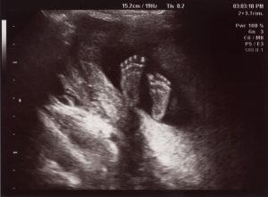
You're now halfway through your pregnancy!
What's happening in my body?
You may have your anomaly scan this week. The sonographer will be checking your baby's development and will also examine your placenta (that's the pancake-shaped organ that feeds your baby and removes waste).
You might find yourself being woken up at night by sudden sharp pains in your calves.
It's probably cramp, which is common in pregnancy and caused by muscular spasms. Rub the muscle hard or pull your toes up towards your ankle.
Exercising more in the day could help you avoid this, and you could try these foot exercises .
Maternity Certificate to claim maternity pay and benefits
Ask your doctor or midwife for a Maternity Certificate, also known as a MAT B1 form. It's issued from 20 weeks onwards.
You'll need it to claim maternity pay and benefits.
Whooping cough jab
Whooping cough is on the rise – but you can protect your baby from this dangerous condition by having a vaccination.
The NHS recommends that all pregnant women should have the jab, ideally between 16 and 32 weeks.
The immunity that you get will be passed on to your baby through the placenta and then offer protection until the routine jab that most babies have at 2 months.
2nd trimester pregnancy symptoms (at 20 weeks)
Your signs of pregnancy this week could include:
- tiredness and sleeping problems ( week 19 has information about feeling tired )
- stretch marks ( read about stretch marks on week 17's page )
- swollen and bleeding gums ( week 13 has information about gum health during pregnancy )
- pains on the side of your belly, caused by your expanding womb (known as "round ligament pains")
- bloating and constipation (read about bloating on week 16's page )
- indigestion and heartburn ( week 25 talks about digestive problems )
- sore breasts
- feeling hot
- swollen hands and feet
- urine infections
- vaginal infections ( see week 15 for vaginal health )
- darkened skin on your face or brown patches – this is known as chloasma or the "mask of pregnancy"
- greasier, spotty skin
- thicker and shinier hair
You may also experience symptoms from earlier weeks, such as:
- morning sickness (read about dealing with morning sickness on week 6's page )
- weird pregnancy cravings (read about pregnancy cravings on week 5's page )
- a heightened sense of smell
- mood swings ( week 8's page has information on mood swings )
- a white milky pregnancy discharge from your vagina and light spotting (seek medical advice for any bleeding)
Read Tommy's guide to common pregnancy symptoms.
What does my baby look like?
Your baby, or foetus, is around 25.6cm long. That's approximately the size of a banana.
Measurements are now taken from head to heel. In earlier weeks, babies are measured from the head to the bottom because the legs are curled up and hard to see.
They are now covered in a white, greasy layer of "vernix". It's thought this protects their delicate skin from drying out in the amniotic fluid. This slippery layer also helps babies to make their way down the birth canal.
Your baby will be getting more active each day. As well as kicking, punching and turning around, your baby could be sucking their thumb – this develops their sucking reflex, which they'll need to feed once they're born.
You may start to feel a bubbling or fluttering in your belly – this could be your baby moving around.
Action stations
You do not have to tell your employer for several more weeks, but as soon as you do, you will have maternity rights and can attend antenatal appointments during paid work time. You can also ask for a risk assessment of your work place to ensure that you're working in a safe environment.
If you want to wait, the latest you can leave it is 15 weeks before the baby is due, which is around week 25.
It's a good time to tone up your pelvic floor muscles. Gentle exercises can help to prevent leakage when you laugh, sneeze or cough.
Get the muscles going by pretending that you're having a pee and then stopping midflow.
Visit Tommy's for more information on pelvic floor exercises.
Ask your midwife or doctor about online antenatal classes – they may be able to recommend one. The charity Tommy's has lots of useful information on antenatal classes and preparing you for birth .
Even if you've had children before, antenatal classes are still worth going to as you can meet other parents-to-be. The NCT offers online antenatal classes with small groups of people that live locally to you.
Do your best to stop smoking and give up alcohol , and go easy on the tea, coffee and anything else with caffeine .
Ask your midwife or GP for support.
To keep bones and muscles healthy, we need vitamin D.
From late March/early April to the end of September, most people make enough vitamin D from sunlight on their skin. However, between October and early March, you should consider taking a daily vitamin D supplement because we cannot make enough from sunlight.
Some people should take a vitamin D supplement all year round, find out if this applies to you on the NHS website.
You just need 10 micrograms (it's the same for grown-ups and kids). Check if you're entitled to free vitamins .
It's recommended that you do 150 minutes of exercise a week while pregnant .
You could start off with just 10 minutes of daily exercise – perhaps take a brisk walk outside. Check out Sport England's #StayInWorkOut online exercises (scroll to the pregnancy section).
Listen to your body and do what feels right for you.
There's no need to eat for 2. You do not need any extra calories until the third trimester, which starts in week 28.
You just need to eat a healthy balanced diet, with a variety of different foods every day, including plenty of fruit and veg. Have a look at our guide to healthy eating in pregnancy .
You may be able to get free milk, fruit and veg through the Healthy Start scheme .
More in week-by-week
As you start the 2nd half of your pregnancy, you'll be entering a period of rapid growth. Your baby is getting ready for life outside the womb and developing essential skills including sucking and breathing.

More in week-by-week guide to pregnancy
Sign up for emails
Our emails include NHS trusted advice and support, tailored to your stage of pregnancy or baby's age.
variable presentation at 20 weeks

IMAGES
VIDEO
COMMENTS
Abnormal Fetal lie, Malpresentation and Malposition
In face presentation, the baby's neck arches back so that the face presents first rather than the top of the head.. In brow presentation, the neck is moderately arched so that the brow presents first.. Usually, fetuses do not stay in a face or brow presentation. These presentations often change to a vertex (top of the head) presentation before or during labor.
Introduction. Cephalic presentation is the most physiologic and frequent fetal presentation and is associated with the highest rate of successful vaginal delivery as well as with the lowest frequency of complications 1.Studies on the frequency of breech presentation by gestational age (GA) were published more than 20 years ago 2, 3, and it has been known that the prevalence of breech ...
Fetal presentation, or how your baby is situated in your womb at birth, is determined by the body part that's positioned to come out first, and it can affect the way you deliver. At the time of delivery, 97 percent of babies are head-down (cephalic presentation). But there are several other possibilities, including feet or bottom first (breech ...
Cephalic occiput posterior. Your baby is head down with their face turned toward your belly. This can make delivery a bit harder because the head is wider this way and more likely to get stuck ...
Negative.No late or significant variable decelerations. Positive.Late decelerations following 50 percent or more of contractions (even if the contraction frequency is fewer than three in 10 minutes).
In breech presentation, the presenting part is a poor dilating wedge, which can cause the head to be trapped during delivery, often compressing the umbilical cord. For breech presentation, usually do cesarean delivery at 39 weeks or during labor, but external cephalic version is sometimes successful before labor, usually at 37 or 38 weeks.
20 Week Scan (Morphology Scan) - Women's Imaging. This is the main ultrasound scan that is obtained during the pregnancy. By 20 weeks most of your baby has developed such that screening of the organs is possible to assess for abnormalities. This scan can be done anytime between 18-22 weeks of your pregnancy but ideally around the 20 week mark.
20 Weeks Pregnant: Baby Development, Symptoms & Signs
Flo for Partners. Anonymous Mode. Find out what窶冱 happening in your body at 20 weeks pregnant, the size of your growing baby, and what symptoms you might experience at this stage.
By week 20, you've probably felt your baby move. It may not feel like an obvious kick—yet! Instead, your baby's first movements might feel like "butterflies" or gas bubbles. Wondering what your baby looks like at 20 weeks? Your baby is now about the size of a red pepper. Average baby length is 6.3 in. (160 mm). How your baby is changing
2 years ago. Asked for Female, 24 Years. Variable presentation during 20 week anomaly scan, is it normal? Does the baby position change with time? 1474 Views v. Answers ( 7) Like the answers? Consult privately with the doctor of your choice. Dr. Lavina Batra.
20 weeks pregnant symptoms. You're in the middle of your second trimester. Your appetite is likely back to normal, or it has increased. Nausea and fatigue may have disappeared during your second ...
These two explanations are based on an Estimated Fetal Weight (EFW) calculations that use the ultrasound technique to evaluate a baby's weight inside its mother's uterus.. To estimate the fetal weight, we use the following measurements:. Abdominal circumference - circumference of the baby's belly, measured at the liver's height.(Advanced: an umbilical portion of the left portal vein should ...
Sonography Fetal Assessment, Protocols, and Interpretation
On March 21, 2014 the following report was presented by ultrasound of the fetuses: the right fetus has variable presentation, normal amniotic fluid, and a posterior placenta at gestational age of 20 weeks and 6 days and the left fetus has variable presentation, normal amniotic fluid, and a posterior placenta at gestational age of 20 weeks and 3 ...
The decrease from the fetal heart rate baseline should be at least 15 beats per minute and should last for at least 15 seconds, but less than 2 minutes. Variable decelerations can be periodic, meaning they are associated with contractions, or they can be episodic and not associated with uterine contractions. [3]
2nd trimester pregnancy symptoms (at 20 weeks) Your signs of pregnancy this week could include: tiredness and sleeping problems (week 19 has information about feeling tired)stretch marks (read about stretch marks on week 17's page)swollen and bleeding gums (week 13 has information about gum health during pregnancy)pains on the side of your belly, caused by your expanding womb (known as "round ...
Fetal Presentation, Position, and Lie (Including Breech Presentation) Key Points | Abnormal fetal lie or presentation may occur due to fetal size, fetal anomalies, uterine structu